Figure 3.

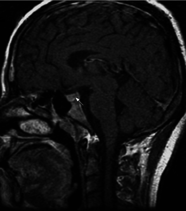
Non-enhanced magnetic resonance image (MRI) of the sagittal section of patient 2 showing the disappearance of the characteristic high signals of the posterior pituitary and increased size of the pituitary stalk.

Non-enhanced magnetic resonance image (MRI) of the sagittal section of patient 2 showing the disappearance of the characteristic high signals of the posterior pituitary and increased size of the pituitary stalk.