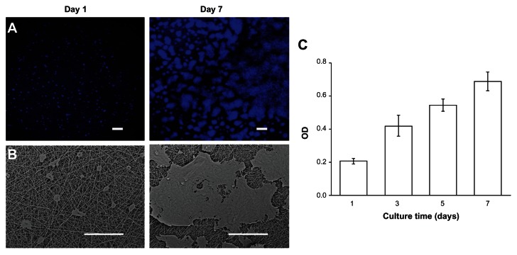
Figure 2.
Biocompatibility of GT/PCL membranes. (A) SEM images of HaCaT cells on a membrane at day 1 and day 7. Scale bars: 200 μm; (B) Confocal microscope images of HaCaT cells on a membrane at day 1 and day 7. Scale bars: 100 μm; (C) Proliferation of HaCaT cells on a membrane measured by a CCK-8 kit.
Abbreviations: GT/PCL, gelatin and polycaprolactone; SEM, scanning electron microscopy; CCK-8, Cell Counting Kit-8; OD, optical density.

