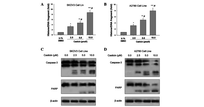
Figure 1.
Effects of casticin on apoptosis in ovarian cancer cells. (A and B) SKOV3 and A2780 cells were treated with the indicated concentrations of casticin for 24 h. Histone/DNA fragmentation was determined using ELISA. Data are presented as mean ± standard deviation (SD; n=3). *P<0.05, **P<0.01 and ***P<0.001 vs. 0.1% dimethyl sulfoxide (DMSO); #P<0.05 vs. treatment with 2.5 μmol/l casticin. (C and D) Cell treatment was the same as in A and B. The expression levels of caspase-3 and poly (ADP-ribose) polymerase (PARP) were determined by western blot analysis in the total cell lysates and β-actin was used as the loading control. ELISA, enzyme-linked immunosorbent assay; SD, standard deviation.

