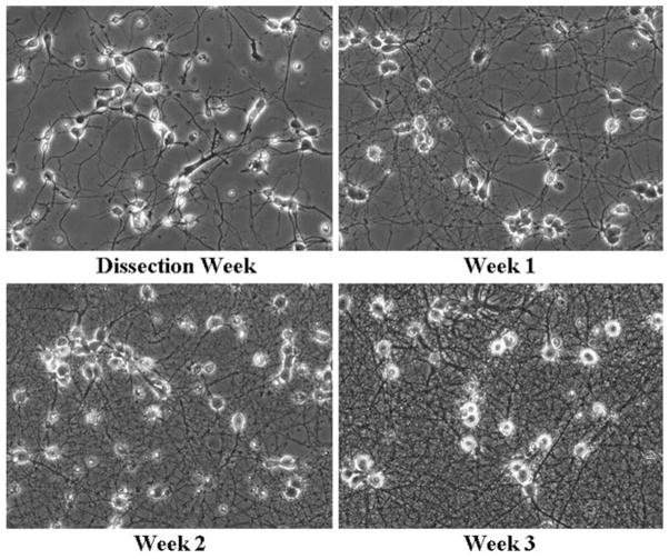
Fig. 3.
Healthy neuronal cultures following dissection. Neuronal architecture was monitored following initial plating until 3 weeks following dissection. Healthy neurons display phase bright somas early on following the dissection with many neurites. Older neurons demonstrate increasing well-defined processes along with phase bright somas.

