
Figure.
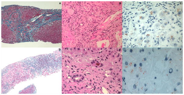
Patient A, liver biopsy at age 14 (A–C): [A] micronodular cirrhosis (Masson trichrome x50); [B] bile duct proliferation without bile retention (arrows) (hematoxylin-eosin x100); [C] hepatocellular copper (Rhodanine stain × 200)
Patient B, liver biopsy at age 6 (D–F): [D] micronodular cirrhosis with severe chronic active inflammation (Masson trichrome x62); [E] portal and lobular inflammation; two small bile ducts (arrows) one with bile plug (hematoxylin-eosin x250); [F] hepatocellular copper (Rhodanine stain x200)