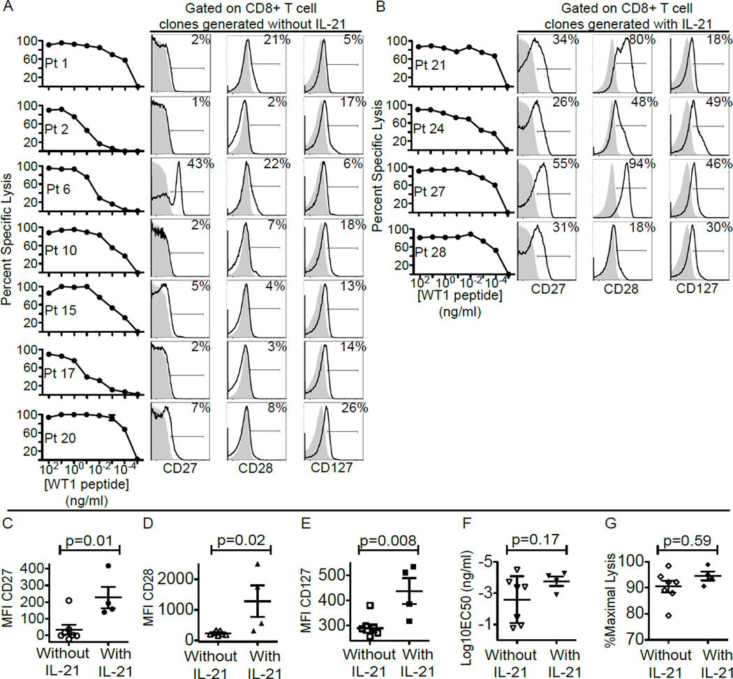
Figure 1. Phenotypic and functional characteristics of WT1-specific CD8+T-cell clones isolated and expanded for infusions.
(A and B) From the left: Lysis by WT1-specific CD8+T-cell clones of TAP-deficient HLA-A*0201+ B-LCL (T2 B-LCL) pulsed with decreasing concentrations of WT1 peptide in a 51Cr-release assay, and expression of CD27, CD28 and CD127 by WT1-specific CD8+T-cell clones (bold line) compared to isotype control (grey area) for clones generated without IL-21 (A) and with exposure to IL-21 (B). Inset values represent percentages of CD27+, CD28+ and CD127+CD8+T-cells respectively. Median fluorescent intensity (MFI) of staining for CD27 (C), CD28 (D), and CD127 (E). Mean effective concentrations of peptide required to achieve 50% lysis (EC50) (F), and percent maximal lysis at an effector to target ratio (E:T) of 10:1 (G) of clones generated without (left) or with IL-21 (right). An unpaired two-tailed equal variance test was used for statistical analysis.

