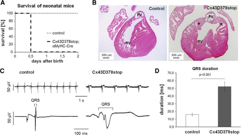
Fig. 1.
Lethality and impaired cardiac conduction in neonatal Cx43D378stop mice. a Homozygous Cx43D378stop:αMyHC-Cre mice die within the first day after birth. In contrast, control littermates (heterozygous Cx43D378stop, heterozygous or homozygous Cx43floxD378stop mice) showed a normal life expectancy after birth. n > 10 per group. b In contrast to controls (left panel) newborn Cx43D378stop hearts (right panel) display irregular thickened trabeculae with abnormal pouch formation (asterisk) at the subpulmonary outflow tract but in contrast to Cx43KO mice the pulmonary outflow tract is still open. PV, pulmonary vein; RV, right ventricle. n > 7 per group. c Representative examples of surface-ECG recordings of a control (left) and a Cx43D378stop (right) fetus (ED 16.5). d Statistical analysis reveal significantly 3.3-fold prolonged QRS intervals in mutant mice compared to controls. n = 6 for Cx43D378stop and n = 7 for control mice

