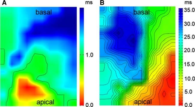
Fig. 3.
Epicardial mapping of Langendorff-perfused hearts from control and Cx43D378stop mice. Representative examples of spontaneous conduction in the left ventricle of a control animal (a) and a Cx43D378stop mouse (b) during sinus rhythm. Isochronal maps show a broad conduction delay in mutant mice. Isochronal lines are shown at distances of 1 ms. n = 3 per group

