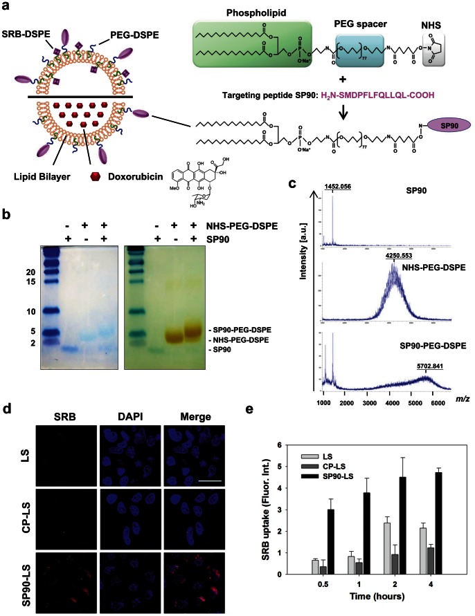
Figure 2. Generation of peptide-conjugated liposomal nanoparticles.
a, Schematic illustration of the targeting liposome, depicting the lipid bilayer membrane encapsulating a large amount of doxorubicin or sulforhodamine B, and the breast cancer targeting ligands that can be displayed on the surface of the liposome. Targeting peptide SP90 was chemically conjugated to NHS-PEG-DSPE in a 1.1∶1 molar ratio. There were about 500 peptide molecules per liposome. b, The percentage of conjugation between SP90 and NHS-PEG-DSPE was confirmed by subjecting the reaction product to electrophoresis on a Tricine-SDS gel, staining with coomassie blue for peptide, followed by barium chloride for PEG (SP90∶1.4 kDa, NHS-PEG-DSPE: 4.3 kDa, SP90-PEG-DSPE: 5.7 kDa). PEGylation efficiency for SP90 was 85% based on quantification of band intensity by densitometry. c, The SP90-PEG-DSPE conjugate was analyzed by MALDI-TOF mass spectrometry (bottom panel). A major peak appears at m/z 5702.8, which can be assigned as the PEGylated SP90 conjugate. Unconjugated SP90 (split peak at m/z 1452) and unreacted PEG (a broad peak around m/z 4250) were also visualized in the mass spectrum, which correspond to the peaks in the top and middle panels, respectively. d, Internalization of SP90-liposomal SRB (SP90-LS) and nontargeted LS (CP-LS and LS) by BT483 cells was studied by confocal microscopy (SRB, red). Nuclear staining was by DAPI (blue) (Scale bar: 10 µm). e, Time-course of SRB uptake by BT483 cells treated with SP90-LS, CP-LS and LS at the indicated times.

