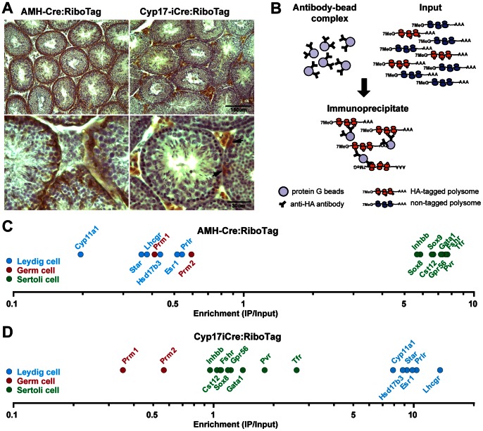
Figure 1. Activation of the RiboTag in Leydig or Sertoli cells of the testis.
(A) RiboTag mice were crossed to a Leydig cell-specific Cre line (Cyp17iCre mice) or a Sertoli cell-specific Cre line (AMH-Cre mice) and the RiboTag activation in the cell type of interest was verified by immunohistochemistry using an anti-HA antibody in testis sections. Arrows point to an unexpected RiboTag activation in scattered cells of the tubule in the Cyp17iCre: RiboTag mice. (B) Cartoon depicting the RiboTag assay. Magnetic beads coupled to anti-HA antibodies were added to testis homogenates of Cyp17iCre: RiboTag or AMH-Cre: RiboTag mice (input of the IP) and incubated overnight at 4°C to capture the HA-tagged polysomes from Leydig or Sertoli cells. After incubation, immunoprecipitates were recovered prior to RNA isolation and analysis. (C) Microarray analysis results confirm the enrichment for well-established Sertoli cell-specific transcripts, as well as the negative enrichment for Leydig and germ cell markers in the IPs from AMH-Cre: RiboTag mice. (D) Similar analysis in Cyp17iCre: RiboTag IPs revealed a significant enrichment for Leydig cell-specific transcripts and a negative enrichment for germ cell-specific mRNAs. Sertoli cell specific transcripts did not show a significant negative enrichment consistent with the RiboTag activation in some Sertoli cells. The enrichment was calculated as the ratio of the signal in the IPs to their inputs.

