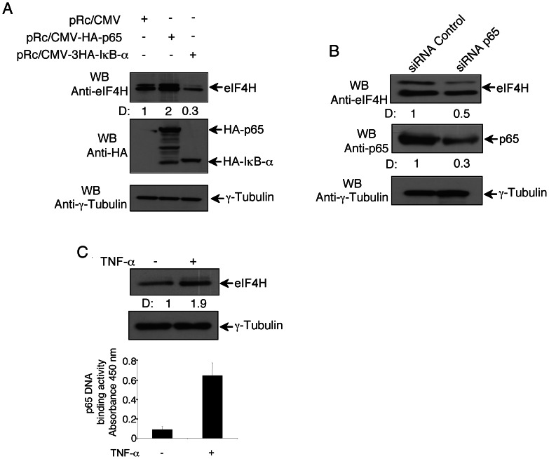
Figure 3. p65-dependent modulation of EIF4H protein expression.
(A) HeLa cells (5×10⁶) were transfected with pRc/CMV-3HA-p65, pRc/CMV-3HA-IκB-α, or pRc/CMV empty vector (5µg), and 48h later whole cell extracts were recovered. Protein extracts (20µg) were separated by 12% SDS–PAGE and analysed by western blotting using anti-HA, anti-eIF4H, or anti-γ-Tubulin antibodies. Densitometry values (D) of the bands were expressed as fold increase above the empty vector, taken as 1. (B) HeLa cells (5×10⁶) were transfected with siRNA control, or siRNA p65 (200 pmol), and forty-eight hours post-transfection whole cell extracts were performed. Protein samples (20µg) were separated by 12% SDS–PAGE, and analysed by western blotting using anti-eIF4H, anti-p65, or anti-γ-Tubulin antibodies. Densitometry values (D) of the bands were expressed as fold increase above siRNA control, taken as 1. (C) HeLa cells (5×10⁶) were 45 min-stimulated with TNF-α (20 ng/mL), or left untreated, washed twice with DMEM, and lysed to perform total extracts and nuclear extracts. Upper panel, total cell extracts (20µg) were separated by 12% SDS–PAGE and analysed by western blotting using anti-eIF4H or anti-γ-Tubulin antibodies. Densitometry values (D) of the bands were expressed as fold increase above un-stimulated cells, taken as 1. Lower panel, nuclear extracts were analysed for the p65 binding to the NF-κB double-stranded oligonucleotide by ELISA EMSA.

