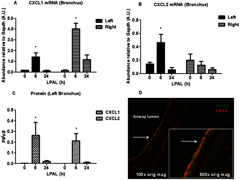
Figure 4. CXCL1 and CXCL2 cytokines mRNA (A, B), protein levels in the left bronchus (C), and (D) frozen section of left bronchus 6 h after LPAL with double staining for CXCL2 (red) and the epithelial cell marker Epcam (green;100× original magnification and inset: 600× original magnification).
CXCL1 (A) and CXCL2 (B) mRNA and CXCL1 and CXCL2 protein levels (C) increased at 6 h LPAL (*P<0.05) and returned to baseline by 24 h LPAL (3–4 rats/time point). Co-localization of stain for epithelial cells (Epcam,;green) and anti-CXCL2 (red) suggest the airway epithelium is a prominent source for CXCL2.

