
Figure 7. Changes in proliferating bronchial vessels.
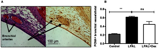
(A) Histologic section of airway demonstrating bronchial vessels by H&E (left panel) and serial section stained for PCNA (right panel). Bronchial vessels show abundant PCNA+ endothelial cells. (B) After LPAL (3 d), a significant increase in the fraction of PCNA+ bronchial vessels is observed. Treatment with dexamethasone had no significant effect on this proliferation index. Control includes both sham and right lungs (3–5 rats/group, **P<0.01 vs control).