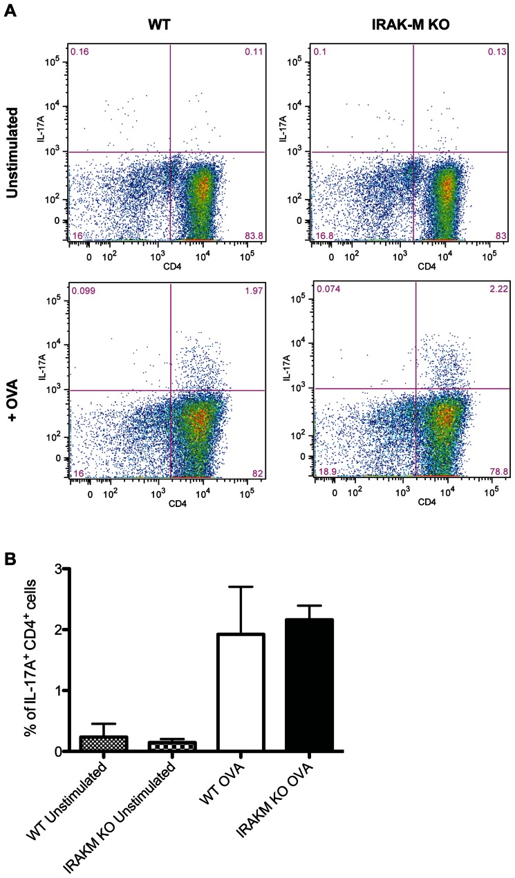
Figure 5. IRAK-M−/− BMDCs do not increase TH17 induction in vitro.
(A) BMDCs isolated from WT and IRAK-M−/− mice were plated and pulsed with OVA for 2 hours before CD4+ T cells isolated from OT-II Foxp3-GFP animals were added to the wells in the presence of IL-6 and TGFβ for 72 hours. Cells were restimulated with PMA and ionomycin in the presence of monesin, and production of IL-17A in CD4+ T cells was measured by flow cytometry. (B) Data are representative of three independent experiments. Bar graph represents mean ± SD from data collected from three individual experiments performed in duplicate.

