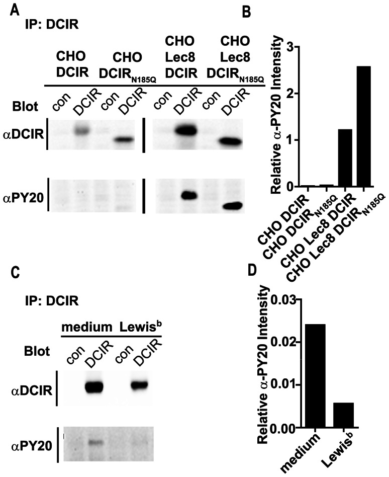
Figure 5. DCIR ligand binding results in decreased phosphorylation of the ITIM in DCIR.
(A) DCIR phosphorylation is decreased in CHO-DCIR cells. Whole cell lysates from DCIR expressing cells were prepared and subjected to immunoprecipitation with α-DCIR (DCIR) or α-Langerin (con, control). Blots were stained for DCIR and for phosphorylated tyrosine (PY20). (B) The relative α-PY20 intensity is calculated by dividing the raw intensity of α-PY20-specific signal in the DCIR immunoprecipitations by the raw intensity of the α-DCIR-specific signal from the corresponding DCIR immunoprecipitations. One experiment out of two independent experiments is shown. (C) DCIR ligand binding results in decreased phosphorylation of the ITIM in DCIR. CHO Lec8-DCIR cells were stimulated with Lewisb. Whole cell lysates were prepared and subjected to immunoprecipitation with α-DCIR (DCIR) or α-Langerin (con). Blots were stained for DCIR and for phosphorylated tyrosine (PY20). (D) The relative α-PY20 intensity is calculated by dividing the raw intensity of the α-PY20-specific signal in the DCIR immunoprecipitations by the raw intensity of the α-DCIR-specific signal from the corresponding DCIR immunoprecipitations. One representative experiment out of three independent experiments is shown.

