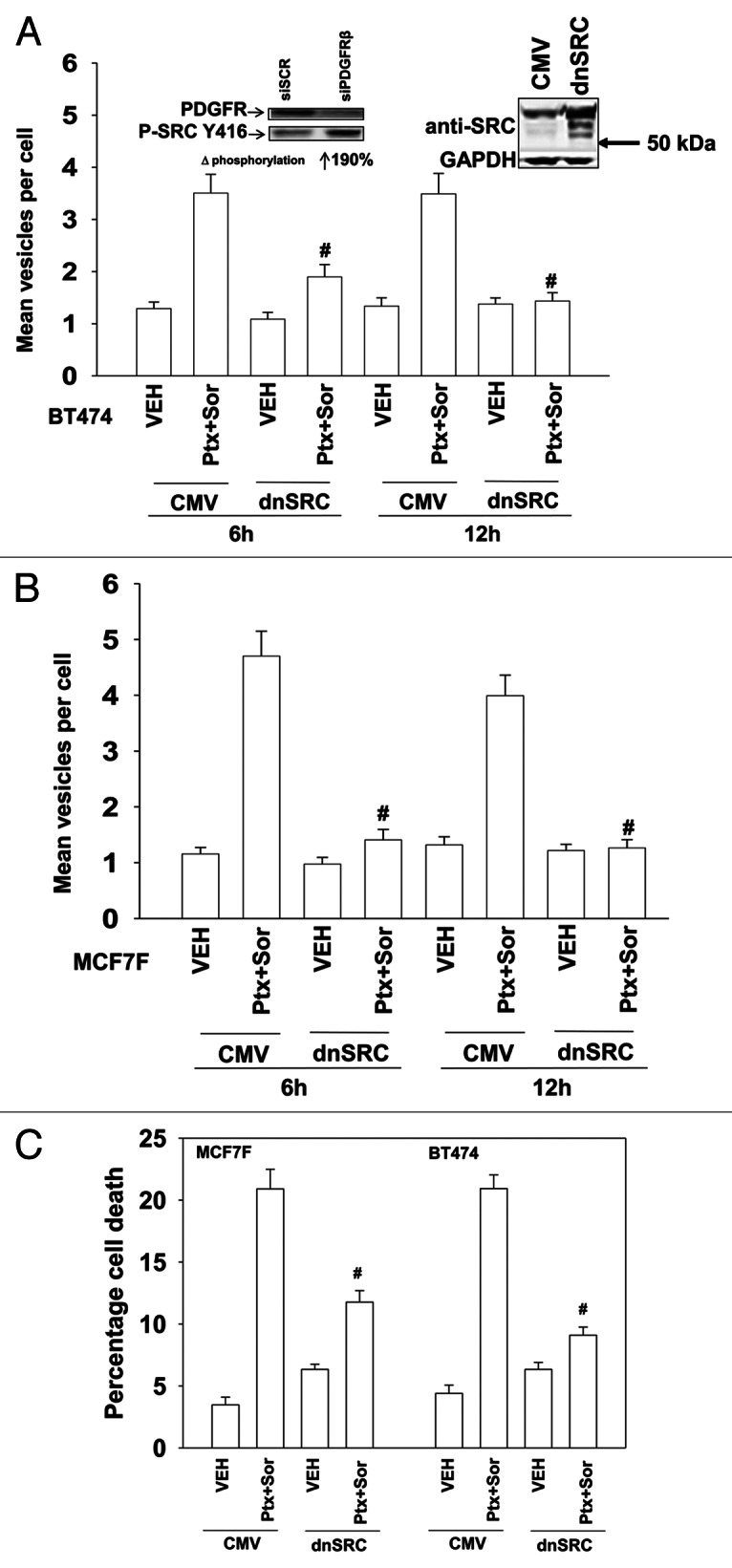
Figure 2. SRC signaling plays an essential role in pemetrexed and sorafenib toxicity. (A) and (B) BT474 and MCF7F cells were transfected to express LC3-GFP and with either empty vector (CMV) or with a plasmid to express dominant negative SRC (dnSRC). After 24h cells were treated with Vehicle (VEH) or pemetrexed (PTX, 1 μM) and sorafenib (SOR, 3 μM), and 6h and 24h later cells were examined under a fluorescent microscope. The mean number of LC3-GFP vesicles per cell was determined (n = 3, ± SEM) # p < 0.05 less than corresponding value in CMV transfected cells. Upper panels: Knockdown of PDGFRβ increases SRC Y416 phosphorylation; increased total expression of c-SRC in cells expressing dominant negative c-SRC. (C) MCF7F and BT474 cells were transfected with either empty vector (CMV) or with a plasmid to express dominant negative SRC. After 24h cells were treated with Vehicle (VEH) or pemetrexed (PTX, 1 μM) and sorafenib (SOR, 3 μM). Cells were isolated 24h later and viability determined by trypan blue exclusion (n = 3, ± SEM) # p < 0.05 less than corresponding value in CMV transfected cells.
