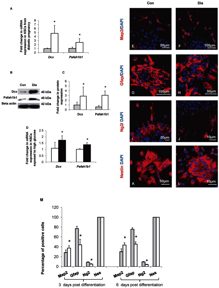
Figure 2. Increased Dcx and Pafah1b1 expression and increased neurogenesis in NSCs from diabetic pregnancy.
(A) mRNA expression of Dcx and Pafah1b1 increased significantly in NSCs from diabetic pregnancy (open bars) compared to the control (filled bars). (B, C) The expression and quantities of Dcx and Pafah1b1 proteins in NSCs from control and diabetic pregnancy were estimated by Western blot. (B) Representative blot shows the expression of Dcx and Pafah1b1 proteins in NSCs from embryos of control and diabetic pregnancy. (C) The quantities of Dcx and Pafah1b1 proteins increased significantly in NSCs from embryos of diabetic pregnancy (open bars) compared to the control (filled bars). (D) Dcx and Pafah1b1 mRNA increased significantly in NSCs exposed to HG in vitro (filled bars) when compared to the control (open bars). (E–L) Neurospheres from embryos of control and diabetic pregnancy were allowed to differentiate for 3 days in vitro and the expression of neuronal (Map2), glial (Gfap, Ng2), and Nestin positive cell populations were determined by immunocytochemistry. (M) The percentage of Map2 positive cells was significantly increased while the percentages of Gfap and Ng2 positive cells were significantly reduced in NSCs from diabetic pregnancy (open bars) on both 3 days or 6 days post differentiation when compared to the control(closed bars). Data is represented as mean ± SD from at least four independent experiments, *p<0.05.

