
Figure 5. PSA analysis of B. fragilis isolates.
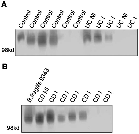
A Western blot of PSA from B. fragilis isolates grown in vitro after isolation from control, CD and UC (inflamed (I) a non-inflamed (NI) biopsies. Panel A: Lanes 1–6 contain B. fragilis isolates from the biopsies control subjects (4, 6, 13, 34, 68, and 1). Lanes 7–8 contain isolates from non-inflamed and inflamed biopsies from subject 23 with UC. Lanes 9–11 contain isolates from inflamed and uninflamed biopsies from subjects with UC: 32, 29 and 77. Panel B: Lane 1 contains B. fragilis type strain 9343 as a positive control. Lanes 2 and 3 contain isolates from a non-inflamed and then an inflamed biopsy from subject 47 with CD. Lanes 4–8 contain B. fragilis isolates from inflamed biopsies of subjects 26, 56, 64, 14, and 85 with CD.