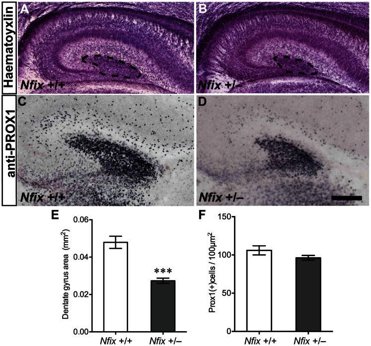
Figure 2. The dentate gyrus is reduced in E18 Nfix +/− mice.
Haematoxylin (A, B) and anti-PROX1 staining (C, D) of E18 wild-type and Nfix +/− coronal brain sections. The overall morphology of the hippocampus in Nfix +/− mice at E18 (B) was normal compared with that of wild-type mice (A), except that the dentate gyrus appeared smaller (dentate gyrus delineated by dashed lines in A, B). PROX1 immunohistochemistry (C, D) confirmed that the overall dentate gyrus area was smaller in Nfix +/− mice at this age (***p<0.001, E). No difference was detected for the number of PROX1-positive cells per unit area of the dentate gyrus (p = 0.19, F). Scale bar (in D): A, B 200 µm; C, D 100 µm.

