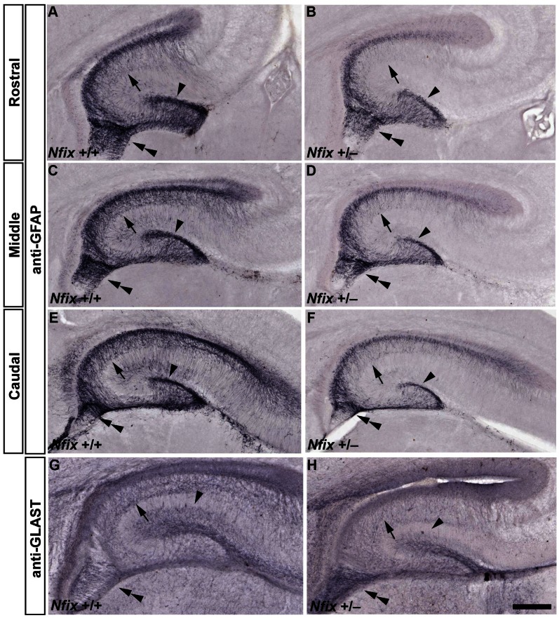
Figure 3. GFAP and GLAST immunoreactivity is reduced in E18 Nfix +/− mice.
Anti-GFAP (A–F) and anti-GLAST staining (G, H) of E18 wild-type and Nfix +/− coronal brain sections. There was reduced GFAP immunoreactivity in the hippocampus of Nfix +/− mice (B, D, F) compared to that of their wild-type littermates at E18 (A, C, E). This reduction in GFAP staining was evident along the rostrocaudal axis of the hippocampus in the supragranular glial bundle (arrowhead), fimbrial glial bundle (double arrowhead) and CA regions (arrow). There was also reduced staining for GLAST in the CA regions and supragranular glial bundle of Nfix +/− mice at E18 (compare panels G and H), although immunoreactivity appeared darker in the fimbrial glial bundle. Scale bar (in H): A–H 250 µm.

