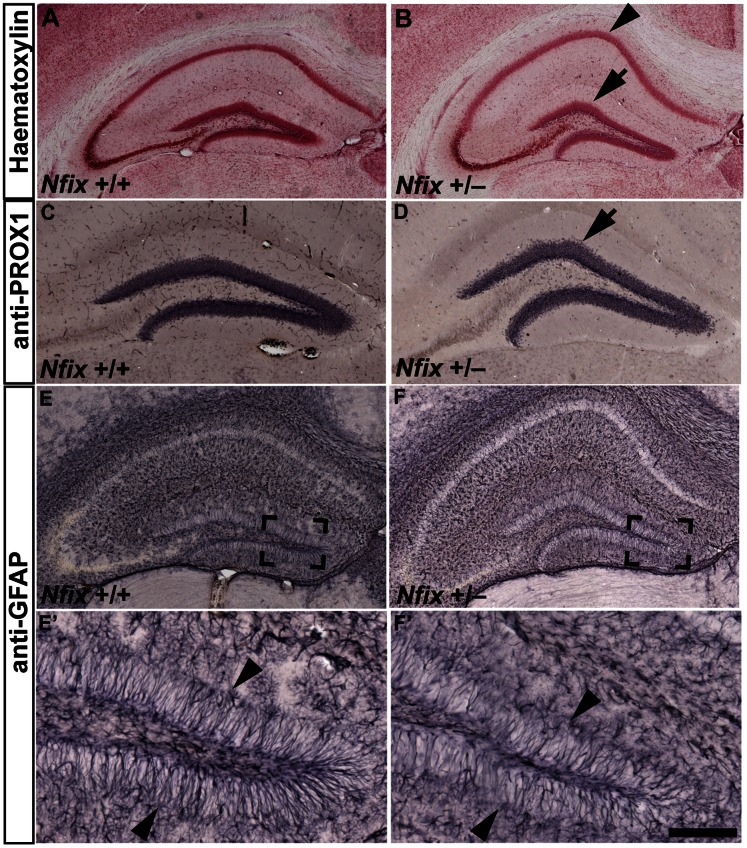
Figure 5. Hippocampal morphology is aberrant in P21 Nfix +/− mice.
Haematoxylin (A and B), anti-PROX1 (C and D) and anti-GFAP staining (E–F’) of coronal sections of P21 wild-type and Nfix +/− brains. In Nfix +/− mice the CA1 pyramidal cell layer had a more pronounced dorsal curvature (arrowhead in B) than in wild-type mice (A). The dentate gyrus was also misshapen in that the lateral region of the suprapyramidal blade met the medial region at a more acute angle (arrow in B and D) than in wild-type mice (A and C). Although no gross difference in GFAP staining was apparent at P21 (E and F), there appeared fewer GFAP-positive radial fibres in the dentate gyrus of Nfix +/− mice (arrowheads in F’) than wild-type mice (arrowheads in E’). E’ and F’ are magnified views of the boxed regions in E and F respectively. Scale bar (in F’): A, B 275 µm; C, D 180 µm; E, F 250 µm; E’, F’ 50 µm.

