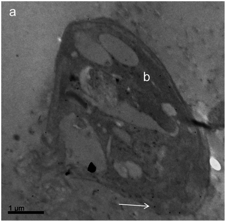
Figure 7. Detection of Pgp 170 molecules, by Transmission Electron Microscopy, after immunogold labeling.
Leishmania amastigote (inside a THP-1 infected cell): cytoplasm of the THP-1 cell (a); Leishmania infantum body (b). Black spots (as indicated by white arrow) show Pgp 170 molecules (gold granules after immunogold labeling using the C219 monoclonal antibody).

