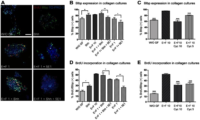
Figure 3. Shh induces tectal NSC proliferation.
Nsp suspension cultures were immobilized in collagen type-I gels. Proliferation experiments were performed for 8 hours and included a BrdU (1 µg/ml) pulse for the last 2 hours in the presence of EGF and FGF-2 (used at 1 ng/ml or 10 ng/ml); combined with either Shh (3,3 µg/ml) or Cyc (5 or 10 µM). (A) Representative images of double immunostaining for BrdU and the RGC marker Blbp after indicated treatments. Bar, 20 µm. (B) Quantification of the percentage of positive cells for Blbp over total numbers of cells, in Z-stacks of confocal microscope. Anti-Shh antibody 5E1 affects both endogenous Shh source as well as recombinant Shh effect resulting in less number of Blbp positive cells. (C) Quantification of the effect of endogenous Shh on the number of Blbp positive cells shows significant inhibition after Cyc treatment. (D) Histogram showing proliferation of nsps after exposures to treatments as indicated. The anti-Shh antibody 5E1 reduces significantly number of BrdU positive cells in the Blbp positive cell population. (E) Quantification of positive cells for BrdU over Blbp positive cells in Z-stacks of confocal microscope treated with Cyc. *, p<0.05; **, p<0.01; ***, p<0.0001. W/O GF: without growth factors, E: EGF, F: FGF-2.

