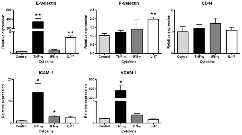
Figure 4.

Graphs showing relative expression of E-selectin, P-selectin, ICAM-1, VCAM-1, and CD44 transcript by immortalized human retinal endothelial cells following exposure to one of the following conditions: medium alone; TNF-α (10ng/ml, R&D Systems, Minneapolis, MN); IFN-γ (20ng/ml, R&D Systems); and IL-17 (100ng/ml, R&D Systems). Endothelial cells were cultured to confluence in modified MCDB-131 medium (Sigma-Aldrich, St. Louis, MO) with 2.5% FBS (Hyclone, Logan, UT) and endothelial growth factors (EGM-2 SingleQuots supplement (Clonetics-Lonza, St. Louis, MO), omitting gentamicin, hydrocortisone and serum, at 1:4 dilution), and subsequently incubated with or without cytokine for 4 hours. Total RNA was isolated using the RNeasy Mini Kit (Qiagen, Valencia, CA), and cDNA was synthesized using the iScript cDNA Synthesis Kit (Bio-Rad Laboratories, Hercules, CA). Relative expression of gene products, normalized to GAPDH, was determined by using the Chromo4 Thermocycler and iQ SYBR Green Supermix (both from Bio-Rad Laboratories). Data were analyzed using Chromo4 Opticon Monitor 3 software. Primer sequences appear in Table 2. In all graphs, bars represent mean and error bars represent standard error of mean (n = 3 wells; * = p<0.05, ** = p<0.01, two-tailed Student’s t-test).
