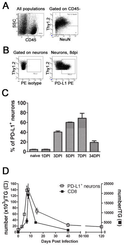
Figure 2. PD-L1/B7H1 expression on neurons in the trigeminal ganglia following HSV-1 infection.

HSV-1 infected TG were excised at various times after infection and dispersed neurons were stained with antibodies against CD45, Thy1.2, PD-L1, and intracellular NeuN. A and B, Representative dot plots showing gating strategies for quantification of PD-L1+ neurons at 8 dpi. C, Bars represent the mean percentage ± SEM of PD-L1+ cellswithin the CD45− Thy1.2+ NeuN+ neuron population (n= 5 mice/time). D, Representative graph shows the average ± SEM number of CD8+ T cells or PD-L1+ neurons in the TG at various times following infection (n = 3–7 mice/time). Three independent experiments produced similar results.
