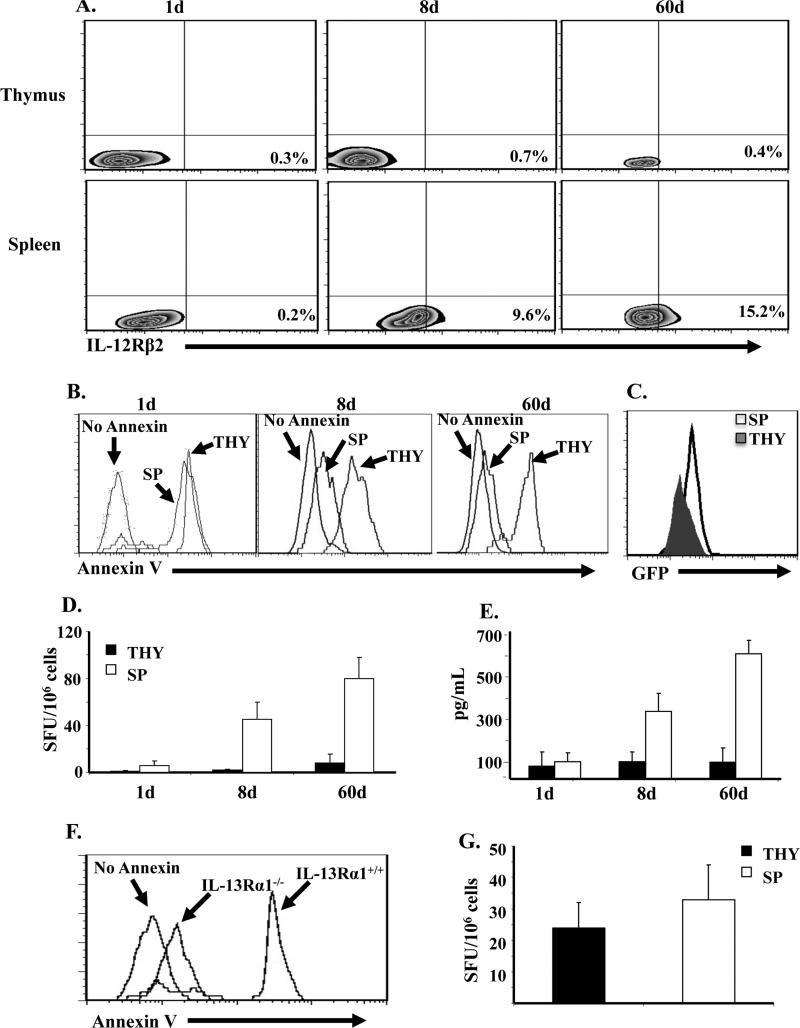
Figure 5. Splenic but not thymic naive CD4 T cells from 8d old mice up-regulate IL-12Rβ2 expression and develop recall Th1 responses upon re-exposure to Ag.
(A) Naïve CD4 T cells were purified from the spleen (SP) and thymus (THY) of DO11.10 mice on day 1 (1d), 8 (8d) and 60 (d60) after birth and assessed for IL-12Rβ2 expression by flow cytometry. The plots show IL-12Rβ2 expression on KJ1-26+CD4+CD62L+ cells. (B-D) Purified SP and THY CD4 T cells from 1, 8, and 60 day-old IL-13Rα1-GFP DO11.10 mice were transferred (105 cells per mouse) into 1d old Balb/c mice and the hosts were given 100 μg Ig-OVA. Two weeks later the SP cells (1× 10⁶ cells/well) were stimulated with 10 μM OVA peptide in vitro and analyzed for Annexin V binding (B), IL-13Rα1 expression (C), and IFNγ production by both ELISPOT (D) and ELISA (E). The histograms show Annexin V binding (B) and GFP expression (C) by cells gated on CD4+KJ1-26+IFNγ+. (D and E) Bars represent the mean ± SD of triplicate wells. (F and G) Purified SP and THY CD4 T cells from 1d DO11.10 IL-13Rα1-/- or IL-13Rα1+/+ control mice were transferred (105 cells per mouse) into 1d old Balb/c newborns and the hosts were given 100 μg Ig-OVA. Annexin V binding (F) and recall IFNγ responses (G) were measured by ELISPOT as described in B and D.

