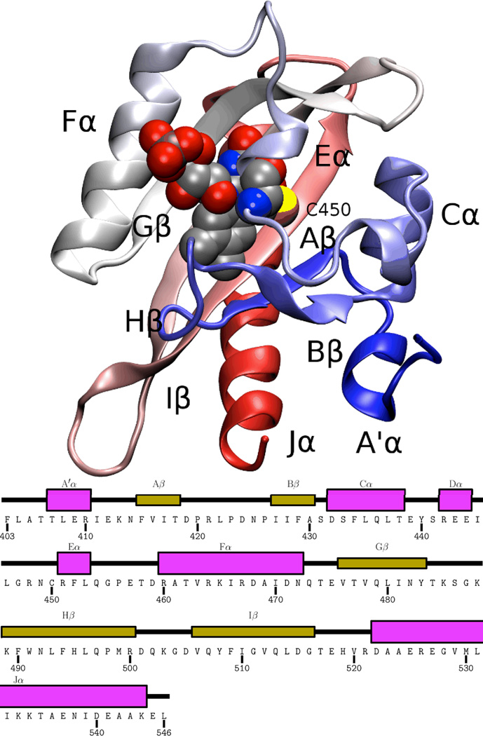
Figure 1.
Overview of the AsPhot1-LOV2 structure 23. The backbone of the homology model is shown in cartoon representation, with the FMN and photoreactive cysteine (C450) shown in vdW representation, and all secondary structure elements labeled. Coloring of the protein backbone runs blue to red from N terminus to C terminus.

