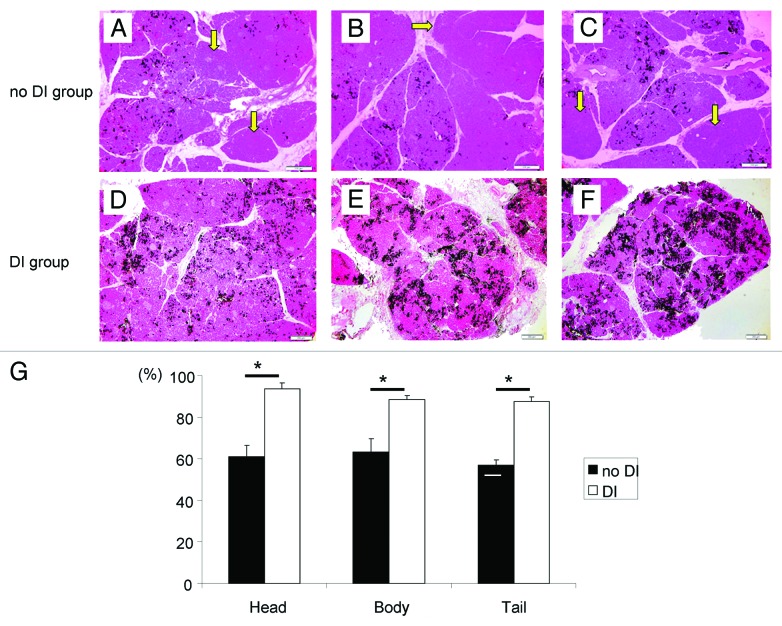
Figure 4. Histologic appearance of the distended pancreas of the no DI group (A–C) and the DI group (D–F) after collagenase solution and 1% dye infusion. In the DI group, dye was found in almost all lobules. However, in the no DI group, the dye was not found in some lobules. Arrows indicate dye-negative lobule (A–C). Magnification: × 40. Scale bars: 200 μm. (G) The ratio of the dye-positive lobules to all lobules in Head, Body and Tail parts of the pancreas. The DI group had a significantly higher rate in all parts. *p < 0.05.

An official website of the United States government
Here's how you know
Official websites use .gov
A
.gov website belongs to an official
government organization in the United States.
Secure .gov websites use HTTPS
A lock (
) or https:// means you've safely
connected to the .gov website. Share sensitive
information only on official, secure websites.
