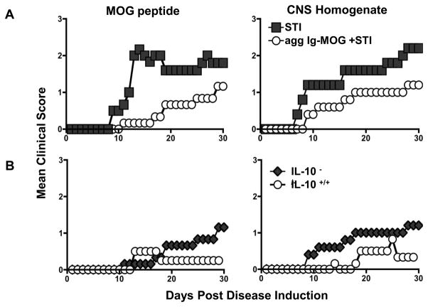
Figure 2. Evaluation of the contribution of IL-10 to Ig-MOG induced suppression of EAE.
(A) IL-10−/ − C57BL/6 mice were induced for EAE with MOG peptide (left) or CNS homogenate (right), and seven days later were treated orally with agg Ig-MOG + STI four times at 2-day intervals. Mice were monitored daily for disease severity for 30 days. The graphs show the mean clinical scores of mice treated with agg Ig-MOG + STI or STI alone. (B) shows comparison of mean clinical scores of IL-10−/ − and IL-10+/+ C57BL/6 mice induced for EAE with MOG peptide (left) or CNS homogenate (right), and treated orally with agg Ig-MOG + STI as in (A). Data is representative of two independent experiments with 5 mice per group.

