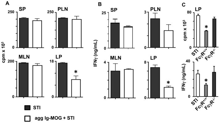
Figure 3. Lamina propria APCs from oral Ig-MOG treated mice display a diminished ability to stimulate MOG specific T cells.

Groups of C57BL/6 mice were given STI alone or 1mg oral agg Ig-MOG + STI and their APCs from different lymphoid tissues were tested for stimulation of MOG-specific 2D2 T cells. (A) shows the proliferative responses of 2D2 CD4 T cells stimulated with MOG peptide-loaded SP, PLN, MLN and LP APC. (B) shows the cytokine responses of the 2D2 T cells described in (A) as measured by ELISA. (C) shows proliferative and IFNγ responses of 2D2 T cells stimulated with MOG peptide-loaded LP APCs that were obtained from FcγR+/+ and FcγR−/ − C57BL/6 mice recipient of 1mg oral agg Ig-MOG+STI. APCs from FcγR+/+ mice treated with STI alone were included for control purposes. Data is representative of two independent experiments and each bar represents the mean ± SD of triplicate wells from 3 to 4 mice. *p < 0.04 (A and B) or < 0.003 for (C).
