Figure 1.
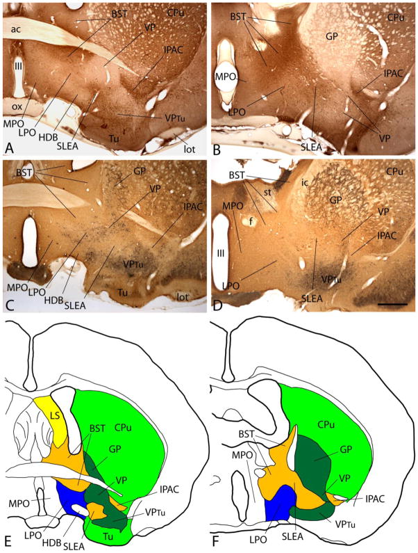
A–D: Photomicrographs illustrating sections through the basal forebrain processed to exhibit nitric oxide synthase (NOS, A and B) or parvalbumin and calbindin-D 28kD (PV and CB, C and D) immunoreactivity (-ir). B and D are cut at a more caudal level than A and B. Note that NOS-ir is strong in extended amygdala stuctures such as the bed nucleus of stria terminalis (BST) and interstitial nucleus of the posterior limb of the anterior commissure (IPAC) and weak in the ventral pallidum (VP). Lateral preoptic area (LPO) exhibits an intermediate density of NOS-ir. PV-ir neurons (black dots) in C and D occupy the VP and, to lesser extent, LPO. E and F: The immunohistochemical patterns in A and B and C and D were used as a basis to draw the maps reflecting the respective levels of the forebrain in E and F. Scale bar: 1 mm for all photomicrographs. Additional abbreviations: ac - anterior commissure; CPu - caudate-putamen; f - fornix; GP - globus pallidus; HDB - horizontal limb of the diagonal band; ic - internal capsule; lot - lateral olfactory tract; LS - lateral septum; MPO - medial preoptic area; ox - optic chiasm; SLEA - sublenticular extended amygdala; st - stria terminalis; Tu - olfactory tubercle; VPtu - ventral pallidum in the deep layer of the olfactory tubercle; III - third ventricle.

