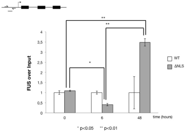
Figure 7. Chromatin immunoprecipitations of CAMK2N2 DNA with FUS and FUS-ΔNLS.
The stable cell lines were induced for six and 48 hours and GFP-tagged FUS-wild type and FUS-ΔNLS were immunoprecipitated using anti-GFP. Chromatin bound to the immunoprecipitates was detected by PCR using the primers located in the promoter regions, as indicated in the cartoon on top. The graph represents three independent experiments. The expression level at time zero was set to one. The differences for FUS-ΔNLS are significant, p<0.05 (*) and p<0.01 (**), as indicated.

