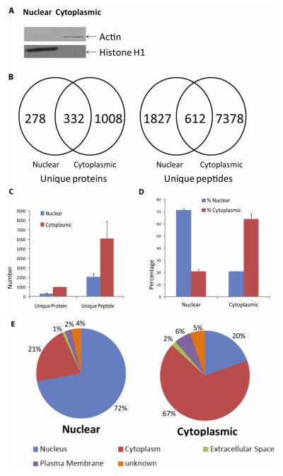
Fig. 1. Comparison of nuclear and cytoplasmic extracts isolated with the basic method.
(A) Western blot detection of cellular compartment-specific proteins in SHSY-5Y cell nuclear and cytoplasmic extracts. Histone H1 (32–33 kD) was found mainly in the nuclear extracts, while actin (42 kD) was enriched mainly in the cytoplasmic extracts. (B) and (C) Comparison of unique proteins and peptides identified from the nuclear and cytoplasmic extracts. Nuclear (Blue bar); Cytoplasmic (Red bar), (B) shows data from one of the representative experiment. Peptide and protein identification criteria are specified in Materials and Methods. Scaffold was used to filter for and compare unique proteins and peptides identified from the analyses of 50 μg of each cytoplasmic or nuclear extract. As expected, more proteins and peptides were discovered in the cytoplasmic extracts. However, 278 unique proteins and 1,827 unique peptides were found only in the nuclear extracts in Fig 1.B (Supplemental Tables 1 & 2). (D) and (E) Cellular localization of the proteins identified from the cytoplasmic and nuclear extracts. (E) shows data from one of the representative experiment. Predictions were made by IPA software. Subcellular localization annotation: Nucleus (Blue); Cytoplasm (Red); Extracellular Space (Green); Plasma Membrane (Purple); Unknown (Orange).

