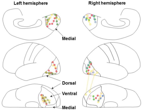
Figure 1. Subdural electrode placement.
The locations of subdural electrodes in 10 patients were superimposed on a brain template (Matsuzaki et al., 2012). The electrodes were implanted on the left hemisphere in five patients and on the right hemisphere in the remaining five (Table 1). The upper panels show the stimulated pairs in the medial-occipital regions; the middle panels show those in the dorsal-occipital regions; the lower panels show those in the ventral-occipital-temporal regions. Each electrode pair is color-coded based on a given patient. All stimulating and recording sites are shown.

