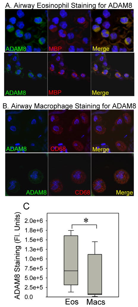
Fig. 10. ADAM8 is expressed at higher levels in eosinophils than macrophages in the lungs of patients with asthma.
Lung sections from an asthmatic subject were double immuno-stained with FITC for ADAM8 (left panels in A and B) and with rhodamine for a marker of eosinophils [major basic protein (MBP) middle panels in A), or with rhodamine for a marker of macrophages (CD68; middle panels in B) and examined using a confocal microscope (original magnification × 600). Merged images are shown in the right panels for A and B. The images shown are representative of immunostained lung sections from three asthmatic subjects. Lung sections stained with non-immune control primary antibodies showed no staining (data not shown). In C, ADAM8 staining in cells identified as eosinophils versus macrophages (by their positive staining with rhodamine using the markers listed above) was quantified using MetaMorph image analysis software. The lines in the boxes in the box plots show the 25th percentiles, medians, and 75th percentiles and the error bars represent the 10th and 90th percentiles. Data are mean ± SEM (n = 18 eosinophils and 9 macrophages). Asterisk indicates p = 0.025.

