
Figure 1. Nuclear EGFR (nEGFR) is detected in early stage NSCLC specimens.
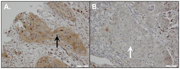
We analyzed 88 primary NSCLC tumors for nEGFR protein expression using immunohistochemistry. (A) Representative case demonstrating nEGFR expression. All positive cases had a similar distinctive pattern of strong nucleolar staining (black arrow). (B) Representative case demonstrating a lack of nEGFR protein expression. Despite the presence of prominent nucleoli, no nEGFR protein is detected (white arrow).