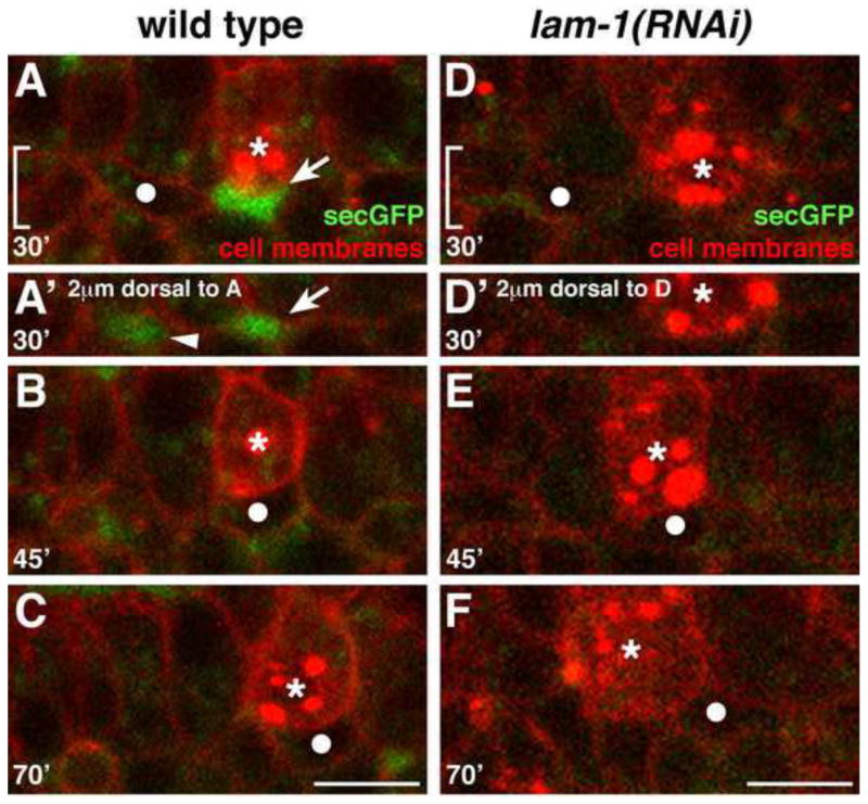
Figure 5. Cell separations are present along the SGP migration route in wild-type but not laminin-depleted embryos.
Stills from a 4D confocal movie of embryo expressing pie-1P::mCherry-PH (cell surfaces), nmy-2P::PGL-1::RFP (PGC-specific P-granules), and pie-1P::secreted GFP (spaces between cells). SGP is indicated with a filled white circle, PGCs with asterisks. Images A–F show a region comparable to that in Fig 1D–F; the midline is at the top of each image. (A–C) Wild-type embryo. (A) Spaces (arrows) can be seen lateral to the endoderm and PGCs, (B–C) and the SGPs migrate into these spaces. (D–F) lam-1(RNAi) embryo. (D–E) Few spaces are evident as the SGPs migrate posteriorly (F) and over-extend past the PGCs. In A′ and D′ the bracketed areas in A and D are shown, at a plane 2μm dorsal to the SGP migration path, to illustrate spaces dorsal to the SGP (arrowhead in A′). Scale bars are 5μm.

