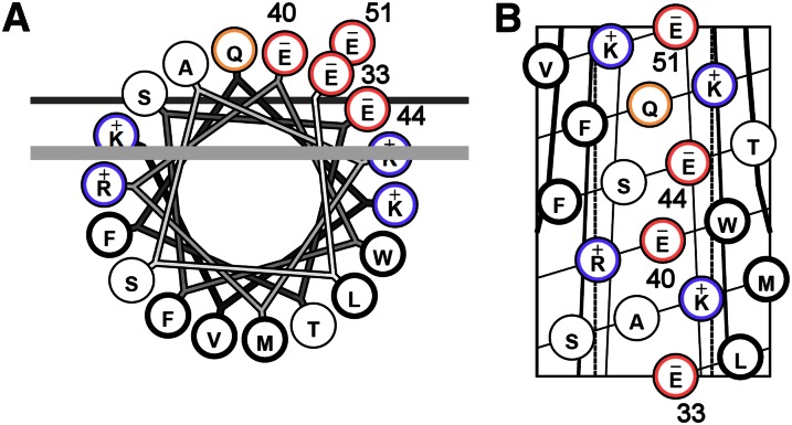
Fig. 1.
Helical wheel projection (A) and helical net (B) representation of apoC-I helix 2 (residues 33–51). (A) Each circle represents an amino acid. The four glutamate residues highlighted in red form a linear acidic array on the polar face of the helix that has been proposed to be required for ABCA1-mediated cholesterol efflux (15). The thick line represents the outside limit of the lipid bilayer in front, and the thin line represents the same in back. (B) Helical net with glutamates numbered. Basic residues are colored blue and acidic residues are colored red. For more information on these helical projections, see Ref. 53.

