Fig. 7.
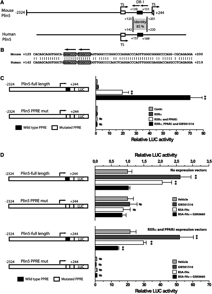
PPARδ regulates Plin5 through a conserved PPRE in intron 1. A: A schematic presentation of the cloned mouse Plin5 reporter (nucleotides −2,324 to +244), and the corresponding human Plin5 promoter. The two alternative transcriptional start sites (TS) are indicated by arrows. The localization of the conserved intron region and percentage identity to the human intronic segment is shown. The nucleotide position of the black boxes points out the localization of the PPREs. B: Sequence alignment and homology between nucleotides in the mouse and the human Plin5 intron 1 regions. The two half sites in the conserved PPRE are enclosed by boxes, and conserved nucleotides are indicated by vertical lines. C: Transient transfection of Plin5 full-length or mutated reporters into C2C12 cells. Cells were transfected, differentiated for 3 days, and stimulated with the indicated ligands for 24 h. Cells were cotransfected with pcDNA3 (control), pcDNA3-RXRα, or pcDNA3-PPARδ expression plasmids and stimulated with vehicle (DMSO) or GW501516 (0.1 μM). Results are presented as mean ± SD (n = 3). D: Transient transfection of Plin5 WT or mutated reporters in combination with no expression plasmid or RXRα and PPARδ expression plasmids. Cells were stimulated with BSA (light gray), GW501516 (0.1 μM; dark gray), BSA-FAs (50 μM OA and 50 μM LA; white), or BSA-FAs in combination with GSK0660 (1 μM; black). For (C) and (D), statistical differences from control treatment (**P < 0.01) or between Plin5 reporter constructs with same treatment (#P < 0.01).

