Figure 1.
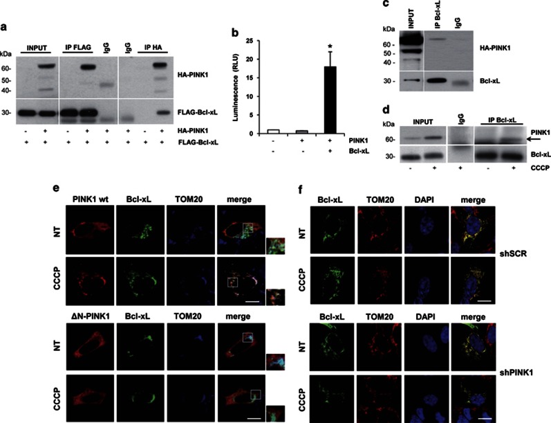
PINK1 interacts with Bcl-xL on depolarized mitochondria. (a) Reciprocal co-immunoprecipitations (co-IPs) of overexpressed PINK1 and Bcl-xL in HEK293 cells. PINK1 and Bcl-xL were immunoprecipitated with HA and FLAG antibodies, respectively. (b) Two-hybrid luciferase assay in HEK293 cells overexpressing PINK1 and Bcl-xL. HEK293 cells were transfected and processed as described in the Method section. In cells overexpressing both PINK1 and Bcl-xL, we observed a significant increase of luminescence compared with negative controls (Relative Light Units (RLU): 17.94±4.03, P=0.026). (c) co-IP of endogenous Bcl-xL and overexpressed PINK1 in SH-SY5Y cells. IP was performed with Bcl-xL antibody, followed by western blotting with HA antibody to detect PINK1. (d) co-IP of endogenous PINK1 and Bcl-xL. Lysates from SH-SY5Y cells, treated with CCCP or vehicle, were subjected to IP with Bcl-xL antibody, followed by immunoblotting with PINK1 antibody. (e) Colocalization of PINK1 and Bcl-xL at mitochondria. SH-SY5Y cells co-transfected with HA-PINK1 wt or ΔN and Bcl-xL–EGFP were treated with 6 h CCCP and subjected to confocal microscopy analysis. PINK1 was immunostained with HA antibody (red); TOM20 antibody was used to label the outer mitochondrial membrane (blue). Bcl-xL strongly colocalized with TOM20 in all experimental settings; conversely, PINK1 wt, but not PINK1-ΔN, colocalized with both Bcl-xL and TOM20 only in the presence of CCCP (see Supplementary Table S1 for overlap coefficients). (f) Colocalization of Bcl-xL and TOM20 upon PINK1 silencing. SH-SY5Y cells stably infected with scramble-shRNA (shSCR) or shPINK1 were transfected with Bcl-xL–EGFP and immunostained with TOM20 antibody (red). Merge pictures reveal colocalization. Bcl-xL strongly colocalized with TOM20 in all settings, regardless of PINK1 silencing (see Supplementary Table S2 for overlap coefficients). Scale bars: 10 μM

