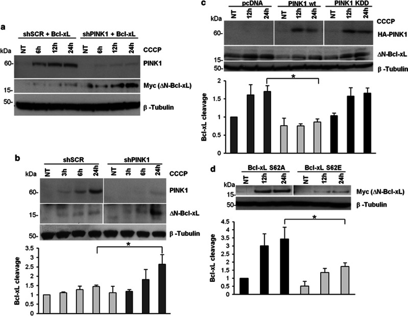
Figure 5.
PINK1-dependent Bcl-xL phosphorylation impairs Bcl-xL cleavage. (a and b) ΔN-Bcl-xL formation upon PINK1 silencing. Lysates from shSCR or shPINK1 cells, either transfected with Myc-Bcl-xL (a) or untransfected (b), were treated with CCCP and processed for western blotting. The cleavage of overexpressed Bcl-xL was well evident upon mitochondrial depolarization in PINK1-silenced cells (a). The endogenous levels of ΔN-Bcl-xL also significantly increased at 24 h CCCP in shPINK1 compared with shSCR cells (2.63±0.52 versus 1.44±0.07, P=0.017). (c) Rescue of Bcl-xL cleavage by PINK1. shPINK1 cells were transfected with PINK1 wt or KDD or vector alone (pcDNA). After 24 h CCCP, cleaved endogenous Bcl-xL was significantly reduced in presence of PINK1 wt compared with both control (0.85±0.09 versus 1.70±0.15, P=0.022) and KDD (1.65±0.14, P=0.022). (d) Cleavage of Bcl-xL S62E and S62A. Lysates from CCCP-treated shPINK1 cells transfected with the indicated Bcl-xL constructs were processed for immunoblotting. Cleaved Bcl-xL was detected using the Myc antibody. At 24 h CCCP, cleavage was significantly reduced in presence of Bcl-xL S62E compared with S62A (1.72±0.23 versus 3.43±0.72, P=0.018). *P-values<0.05

