
Fig. 2.
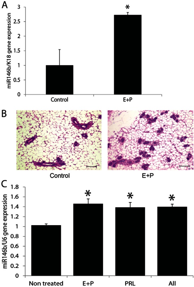
In vivo administration of estrogen and progesterone resulted in upregulation of miR-146b expression in the mammary epithelial cells. (A) Quantitative PCR of miR-146b in PMECs from virgin mice treated with estrogen and progesterone for 3 weeks compared with non-treated virgin mice. Data are means ± s.e.m. (n = 4, *P<0.05). (B) Representative Hematoxylin and Eosin-stained sections from the hormone-treated and control mice. Scale bars: 100 µm. (C) Quantitative PCR of miR-146b in PMECs from virgin mice treated with estrogen plus progesterone (E+P), with prolactin (PRL) or both for 10 days in vitro compared with non-treated PMECs. Data are means ± s.e.m. (n = 3, *P<0.05 compared with the control).