Fig. 7.
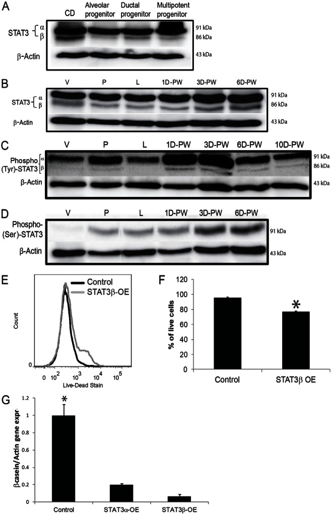
STAT3β is activated in the mammary epithelial cells during involution. (A) Western blot analysis of total STAT3α/β in the CDβ cell line and the CD-derived clones. (B–D) Western blot analysis of total STAT3α/β (B); phosphorylated tyrosine 705 (phospho-Tyr-STAT3; C); phosphorylated serine 727 (phosphor-ser-STAT3; D) in total glands from virgin (V), pregnant (P) and lactating (L) mice and from mice at different physiological stages of involution: days 1, 3 and 6 (B–D) and 10 (C) days post-weaning (D-PW). (E) Representative histogram of flow cytometry analysis of the alveolar progenitor cell line stained with the LIVE/DEAD® Fixable Dead Cell Staining Kit at 24 hours post-transfection with STAT3β overexpression clone. (F) Quantification of the percentage of live cells (n = 3, P<0.05). (G) RT-qPCR of mouse mammary epithelial cells transfected with STAT3α(STAT3α-OE)- or STAT3β (STAT3β-OE)-overexpression clone compared with cells transfected with the empty vector (control). The cells were cultured on Matrigel for 72 hours, with prolactin treatment. Cells were recovered and analyzed for β-casein expression by qPCR (n = 3, *P<0.05 compared with other cells). Data are means ± s.e.m.

