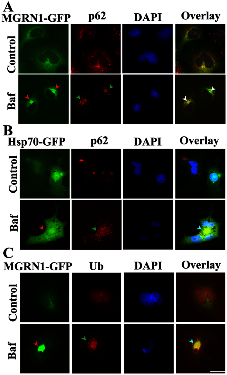
Figure 3. Co-localization of MGRN1 with cytoplasmic p62 bodies, and ubiquitin (Ub) following autophagy dysfunction.
(A) Cos-7 cells were plated on two-chamber slides. On the following day, the cells were transiently transfected with the MGRN1-GFP construct. After 36 h of transfection, the cells were treated with 50 nM Bafilomycin (Baf) for 12 h. Post-treatment, the cells were fixed and subjected to immunofluorescence staining using an p62 antibody. A rhodamine-conjugated secondary antibody was used to label the p62 antibody. Nuclei were stained with 4′,6- diamidino-2-phenylindole (DAPI). Arrows indicate the recruitment of MGRN1 to p62 aggregates. (B) Cells were transfected with Hsp70-EGFP and treated with Baf as described in A. Cells were observed using a fluorescence microscope and their nuclei were stained using DAPI. Arrows indicate the co-localization of Hsp70 with p62 aggregates. (C) Cells were transiently transfected with the MGRN1-GFP construct and treated with Baf as described in A; after treatment, the cells were processed for immunofluorescence staining using ubiquitin (Ub) antibody. Rhodamine-conjugated secondary antibody was used to label ubiquitin. Nuclei were stained with DAPI. Arrows indicate the redistribution of MGRN1 with Ub-positive aggregates. Scale bar, 20 μm.

