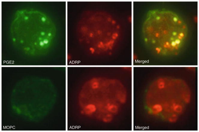
Fig. 10.2.
EicosaCell for PGE2 immunolocalization within BCG-infected cytospun macrophages. In upper panels, macrophages from BCG-infected animals were labeled for ADRP-associated lipid bodies (red staining) and for newly formed PGE2 (green staining). Merged image showed co-localization of PGE2 in ADRP-associated lipid bodies (yellow staining). In bottom panels, IgG1 irrelevant isotype (MOPC) was used as control for PGE2 labeling. Briefly, pleural macrophages obtained 24 h after infection with BCG were recovered from the thoracic cavity with 500 μL of HBSS, immediately mixed with 500 μL of EDAC (1% in HBSS) and incubated for 30 min at 37°C. Cells were then washed with HBSS, cytospun onto glass slides and incubated with mouse anti-PGE2 (1/100) or MOPC 21 in 0.1% normal goat serum and guinea pig polyclonal anti-mouse ADRP (1/1000) in 0.1% normal donkey serum simultaneously for 1 h at RT. Cells were washed twice and incubated with secondary Abs, goat anti-mouse conjugated with AlexaFluor-488 (1/1000) and CY3-conjugated donkey anti-guinea pig (1/1000). The slides were washed (three times, 10 min each) and mounted with aqueous mounting medium.

