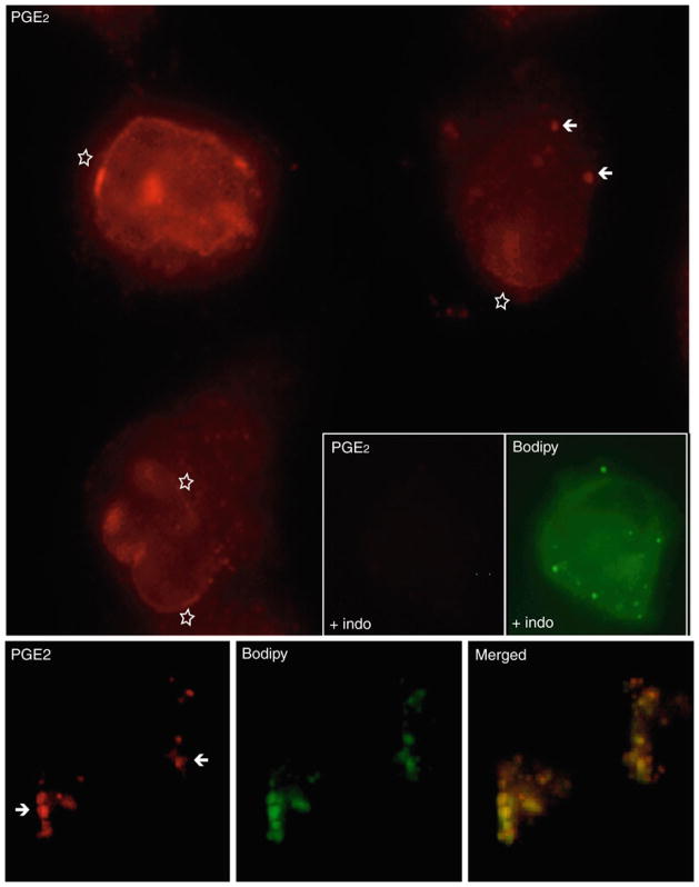
Fig. 10.3.
EicosaCell for PGE2 immunolocalization within adherent CACO-2 cells. The largest panel shows fluorescent microscopy of CACO-2 cells labeled for newly formed PGE2 (red staining). Bottom three images panel showed immunofluorescent PGE2 (red staining), BODIPY-associated lipid bodies (green staining) and a merged image showing co-localization of PGE2 in lipid bodies (yellow staining). Insert panel showed lack of PGE2 immunolabeling within lipid-body-enriched CACO-2 cells, which were treated with indomethacin (4 mg/kg) 1 h before EDAC. Briefly, CACO-2 cells were fixed and permeabilized during 1 h at 37°C with EDAC (0.5% in HBSS−/−). Then, cells were washed with HBSS and blocked with 2% donkey serum for 15 min before incubation with anti-PGE2 monoclonal antibody (Cayman Chemicals) for 45 min. Cells were washed with HBSS and incubated with fluorescent secondary antibody Cy3-conjugated affiniPure F(ab′) fragment donkey anti-mouse and BODIPY 493/503 (Molecular Probes, CA) for 45 min.

