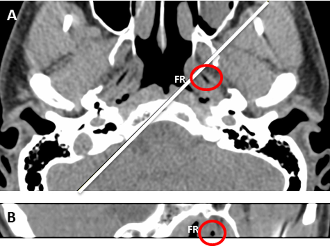
Figure 5.

Computed tomography reconstruction in subject with patulous Eustachian tubes (subject 7). The red circle surrounds the air bolus present in the Eustachian tube. The Fossa of Rosenmuller (FR) can also be seen in both planes. The white line in 5A represents the plane shown in 5B.
