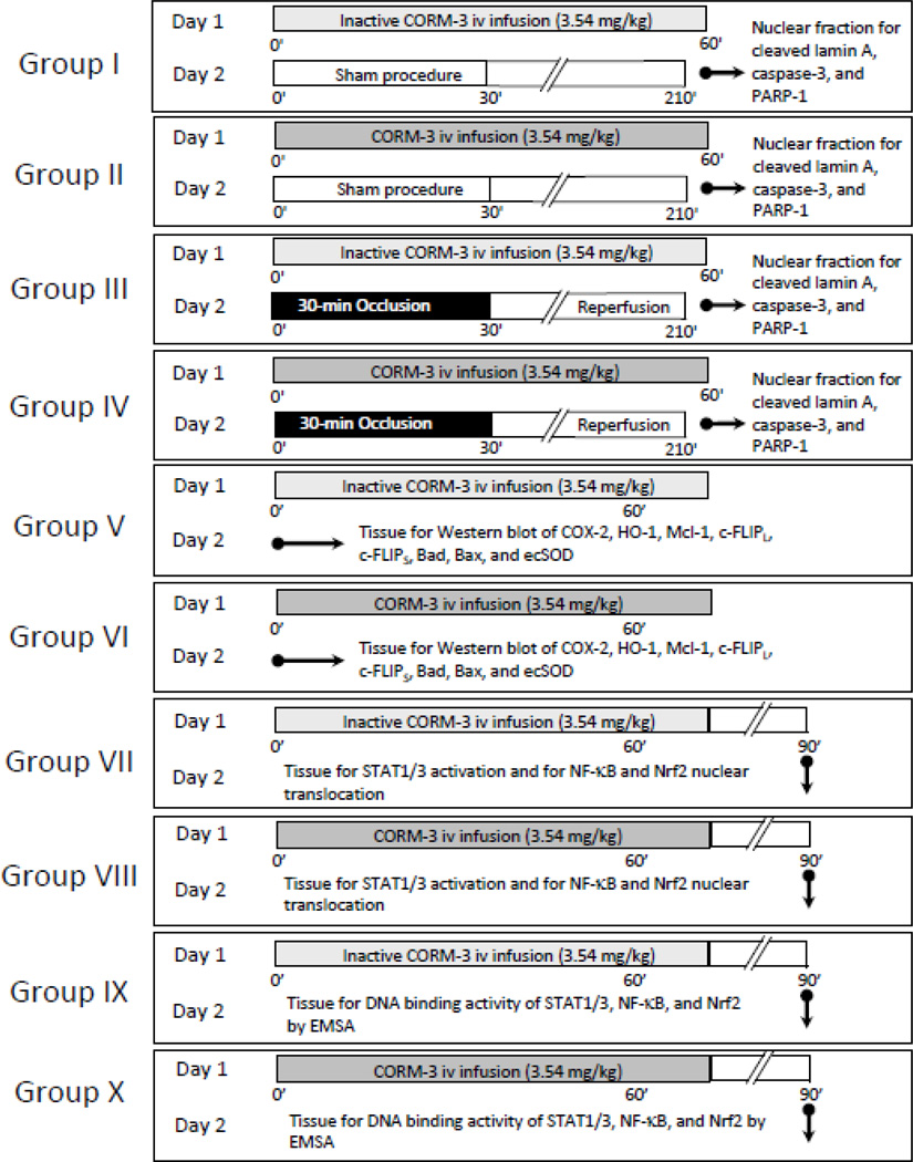
Figure 1.
Experimental protocol. Ten groups of mice were used. On day 1, mice in groups I and III received 3.54 mg/kg of iCORM-3 i.v. over 60 min, while mice in groups II and IV received 3.54 mg/kg of CORM-3 i.v. over the same period. Twenty-four hours later, mice were subjected to either a sham open-chest procedure (groups I and II) or a 30-min coronary occlusion followed by reperfusion (groups III and IV). Three hours later tissue samples were harvested from the risk region for cleaved lamin A, cleaved caspase-3, and cleaved PARP-1 assays. On day 1, mice in groups V and VI received the same dose of iCORM-3 and CORM-3, respectively, and myocardial tissue samples were harvested for the measurement of Mcl-1, c-FLIPS, c-FLIPL, COX-2, HO-1, Ec-SOD, Bad, and Bax 24 h later. For transcription factor assays, mice in groups VII and IX received the same dose of iCORM-3, while mice in groups VIII and X received the same dose of CORM-3. Thirty min after the completion of infusion, myocardial tissue samples were harvested for the determination of nuclear translocation and/or phosphorylation of p65, pTyr-STAT1, pTyr-STAT3, and Nrf2 (groups VII and VIII) and DNA binding activity using gel shift assays (groups IX and X).

