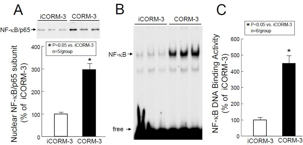
Figure 8.
Nuclear translocation of p65 subunit and NF-κB DNA binding activity. Myocardial tissue samples were harvested from mice in groups VII and IX (iCORM-3-treated) and VIII and X (CORM-3-treated) 30 min after the completion of infusion. (A) Upper panel: A representative Western immunoblot showing increased nuclear levels of p65 in CORM-3-treated hearts compared with iCORM-3-treated hearts. Lower panel: Densitometric analysis of p65 signals. (B) EMSA showing NF-κB DNA binding activity. (C) Densitometric analysis shows a marked increase in NF-κB DNA binding activity in CORM-3-treated hearts compared with iCORM-3-treated hearts. Data are means±SEM. *P<0.05 vs. iCORM-3.

