Figure 2.

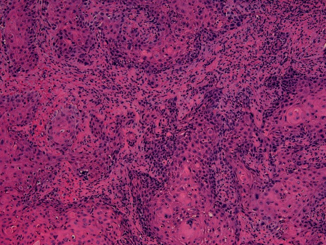
Part of the tumor showed features of conventional squamous cell carcinoma (hematoxylin and eosin stain, 20× magnification).

Part of the tumor showed features of conventional squamous cell carcinoma (hematoxylin and eosin stain, 20× magnification).